개의 항문직장 평활근육종(Anorectal Leiomyosarcoma) 증례
Anorectal Leiomyosarcoma in a Dog
Sohee Lim, Sangjune Sohn, Woo-Jin Song, Yeonjung Hong*. J Vet Clin 2025;42(1):31-36. CC BY-NC 4.0 · KSVC Endorsed Learning Hub
https://doi.org/10.17555/jvc.2025.42.1.31 ↗
16세 중성화 수컷 푸들에서 경막외하부(perineal) 부위에 큰 단단한 종괴가 있어 Sangjune Sohn1,2,† 내원하였다. 컴퓨터단층촬영(CT)상으로는 결장과 직장의 교차부에서 원인으로 추정되는 다발성 석회화 소견을 동반한 거대한 부드러운 조직으로 구성된 종괴가 관찰되었다. 이 종괴는 외상성 절제(surgical excision)를 통해 완전히 제거되었으며, 그 기원은 근위 결장과 회음근(anal sphincter)이었다. 면역조직화학은 신생물세포가 알파 평활근액틴에 양성이고 c-Kit에 음성을 보였으며, 조직병리 및 면역조직화학은 분화가 잘 된 평활근육종(원발 부위: 항문경부)임을 확인하였다. 이 개 사례는 수술 후 1년간 재발 없이 좋은 예후를 보였으며, 유의한 합병증 없이 치료 효과가 나타났다. 또한 이 연구는 현재까지 공개된 자료로 보면 개의 항문직장 평활근육종(anorectal leiomyosarcoma) 사례 중 컴퓨터 단층 촬영(CT) 검사 결과를 포함한 첫 번째 보고서이다. CT 영상검사는 항문 종괴의 크기, 범위, 전이 여부를 사전에 정확히 평가할 수 있게 해서 수술 계획 수립과 예후 평가에 유용한 정보를 제공하였다.
A 16-year-old neutered, male poodle presented with a large, hard Sangjune Sohn1,2,† mass in the dorsal perineal area. Computed tomography revealed a massive Woo-Jin Song2 soft-tissue attenuated mass with multiple calcifications located in the anus, comYeonjung Hong1,* pressing the rectum. The huge mass, originating from the anal sphincter and distal part of the rectum, was completely surgically removed. Immunohistochem- 1 Western Animal Medical Center, Seoul istry revealed that neoplastic cells were positive for alpha-smooth muscle actin 04101, Korea 2 and negative for c-Kit. Histopathology and immunohistochemistry confirmed Laboratory of Veterinary Internal Medicine, College of Veterinary Medicine, well-differentiated leiomyosarcoma originating from the anal sphincter. The paJeju National University, Jeju 63243, Korea tient has shown a good treatment outcome and prognosis, with no significant postoperative complications or recurrence since the surgery during the 1-year follow-up period to date. To our knowledge, this is the first published report of a canine anorectal leiomyosarcoma that includes a computed tomographic investigation. CT imaging enabled successful preoperative assessment of the size, extent, and metastatic status of the anal mass, proving valuable for surgical planning and prognosis evaluation.
최종 진단
조직병리에서 종괴는 불분명한 세포 경계의 방추세포로 구성되었고, 소용돌이형·공포성·호산성~양친화성 세포질을 보였습니다. 핵의 크기·모양 변이(anisocytosis/anisokaryosis)는 경도~중등도였고, 유사분열 수는 10 고배율 시야당 1개(2.37 mm²), 괴사는 약 25%를 차지했습니다.
면역조직화학에서 c-kit 음성, 평활근 액틴(SMA) 양성으로 위장관기질종양(GIST)과 감별되었으며, 절제연 음성의 고분화 평활근육종(well-differentiated leiomyosarcoma)으로 확진되었습니다.
면역조직화학에서 c-kit 음성, 평활근 액틴(SMA) 양성으로 위장관기질종양(GIST)과 감별되었으며, 절제연 음성의 고분화 평활근육종(well-differentiated leiomyosarcoma)으로 확진되었습니다.
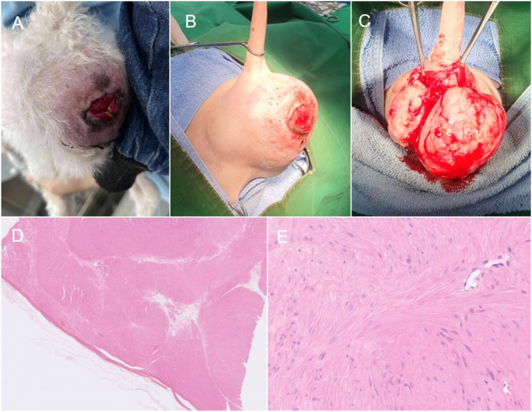 J Vet Clin 2025;42(1) Fig.3 (D,E)H&E (배율 원문 미보고)
조직병리: 얇게 가성피막에 싸인 다결절성, 경계가 불분명한 중등도 세포충실성 종양으로, 불분명한 경계의 방추세포로 구성됩니다.
문제
평활근육종과 GIST를 감별하는 면역조직화학 소견으로 옳은 것은?
해설 정답 b: GIST는 카할 간질세포 기원으로 c-kit(CD-117)을 발현하지만 평활근육종은 c-kit 음성·평활근 액틴(SMA) 양성입니다. 본 증례도 c-kit 음성·SMA 양성으로 확진했습니다. a는 양성 표지를 뒤바꾼 함정, c는 감별 불가라는 오개념, d는 SMA 양성인 사실과 반대입니다.